Fluorine 18 (F 18) is a radioisotope of the element fluorine. This positron-emitter plays a key role in positron emission tomography (PET), three-dimensional, colour imaging of biological processes in the body. When biologically active tissues in the body absorb a solution containing F18, the PET machine detects the gamma rays produced from the collision of positrons emitted from F 18 and nearby electrons. Computer analysis of the gamma ray data produces detailed, multicoloured images of the body at work.
Cancer Diagnosis and Treatment
PET scans are used widely for cancer detection and the monitoring of cancer treatments. Through an IV drip or injection, patients take a solution containing a radioisotope called a radiopharmaceutical. Fluordeoxyglucose (FDG), the most commonly used radiopharmaceutical, is a glucose substitute that contains F 18. Active cells absorb glucose for energy. PET scanners record and analyse the gamma rays produced by positron emissions of the F 18. Since cancer cells are more active than surrounding healthy cells, they absorb more FDG and emit more gamma rays. PET scans precisely distinguish levels of gamma ray activity and translate that data into detailed images of the body's tissue.
Other F18 radiopharmaceuticals allow doctors to hone in on specific disease activity in the body. For example, PET scans using F18 sodium fluoride provide detailed images of bone malignancies, enabling detection of bone cancer and the monitoring of the effectiveness of treatment. F18 is attached to a synthetic peptide to create a solution that images tumour cells and tumour vasculature.
- PET scans are used widely for cancer detection and the monitoring of cancer treatments.
- For example, PET scans using F18 sodium fluoride provide detailed images of bone malignancies, enabling detection of bone cancer and the monitoring of the effectiveness of treatment.
Cognitive Science
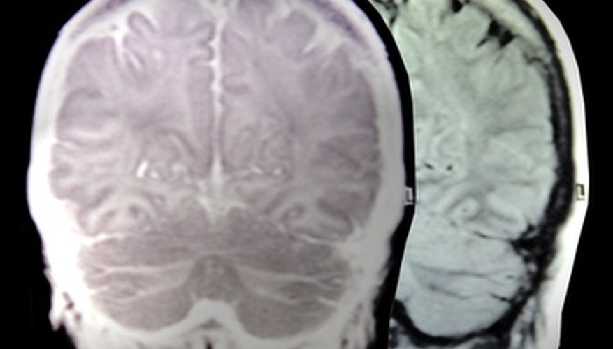
In cognitive research, PET scans help reveal the function or dysfunction of the brain's activity. Researchers can compare images of healthy brains to those of patients with mental disease to gain better understanding of a disease's impact on brain activity. PET scans reveal what parts of the brain work as a person reads, dances, memorises data, eats, or gets scared. F18 makes these scans possible because of its relatively long half-life. Subjects have time to complete an activity while the solution is still actively emitting positrons.
- In cognitive research, PET scans help reveal the function or dysfunction of the brain's activity.
- Subjects have time to complete an activity while the solution is still actively emitting positrons.
Alzheimer's Disease
Researchers at Avid Radiopharmaceuticals in Philadelphia are using F 18 in a new tracer solution, florbetapir, that enables the PET imaging of plaques in the brain associated with Alzheimer's disease. Announced in the New York Times on June 23, 2010, this breakthrough means that doctors can now definitively diagnose Alzheimer's in living patients. Before the breakthrough, definitive diagnosis required an autopsy. In addition to accurate diagnosis, doctors will be able to use the PET scans to monitor the effectiveness of treatments for the disease.
- Researchers at Avid Radiopharmaceuticals in Philadelphia are using F 18 in a new tracer solution, florbetapir, that enables the PET imaging of plaques in the brain associated with Alzheimer's disease.